Physical Therapy
Neuroanatomy
Welcome to Neuroanatomy! 🧠
In light of the evolving situation with COVID-19, we have compiled these online resources to help supplement your learning in lieu of in-person neuroanatomy labs.
For best results, use in conjunction with your favourite anatomy atlas!
Lab 1: Skull, Meninges, & Venous Sinuses
1. Skull (Exterior, Interior, & Inferior)
All bones of the human adult skull—with the exception of the mandible (lower jaw)—are joined by synarthroses (immovable joints) called cranial sutures.
Let’s start with this model of the exterior skull by Aaron Cohen-Gadol M.D. of The Neurosurgical Atlas. Click on “?” at the bottom of the video to view instructions for manipulating 3D models in SketchFab.**
(i) Consider how the bones articulate with each other
(ii) Identify each bone and its prominent features listed in your lab manual (Section A)
(iii) Take note of the main exterior sutures and determine which bones join to form them
**Click “Select an annotation” at the bottom of the video, then click “Show annotations” at the top of the drop down menu to view the complete set of 26 labels.
If, like me, you find it challenging to wrap your head around how the bones of the skull articulate, check out this “Exploding Skull” CT-scanned 3D model by the Witmer Lab at Ohio University College of Osteopathic Medicine. Pause the video at any point during the brief animation. You can then manipulate the model to view the disarticulated bones of the skull from any angle.
Next are 2 skull models from teams at the University of British Columbia Faculty of Medicine.
Start with the first model and refer to Section B and Section C in your lab manual for lists of bony features and foramina that you will need to be able to identify from internal and external (inferior) views, respectively.
(i) Locate the anterior, middle, and posterior cranial fossa. Which bones form each fossa and which foramina are found in each?
(ii) Consider the arrangement of the foramina in the skull. What structure(s) might you expect to exit or enter the skull through each? (this will be reviewed in future labs)
(iii) Identify the bone (or bones) in which each foramen is located
The second model (created by Connor Dunne using Reality Capture photogrammetry at UBC HIVE) includes 13 clickable annotations for your review. Feel free to explore the associated external links to videos and resources on neuroanatomy.ca & clinicalanatomy.ca.
If you’re finally starting to get pǝʇuǝᴉɹosᴉp from manipulating all these 3D models, here are some more traditional illustrations & figures. These are all labelled, but we will provide some links to unlabelled resources & interactive quizzes so you can assess your knowledge of the material on your own time!
These illustrations of the bony skull are borrowed from the 14th edition of Grant’s Atlas of Anatomy (Agur & Dalley) with permission from Dr. Anne Agur. Click each image to enlarge.
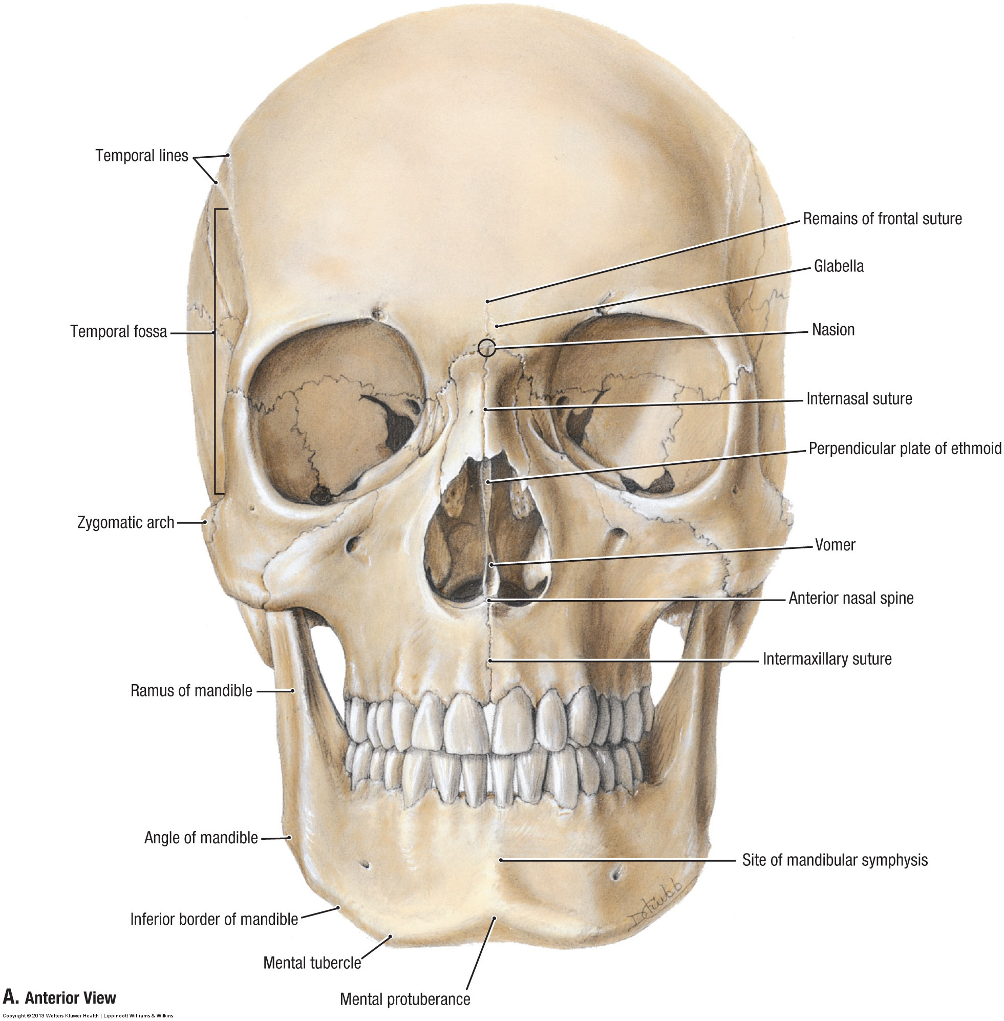
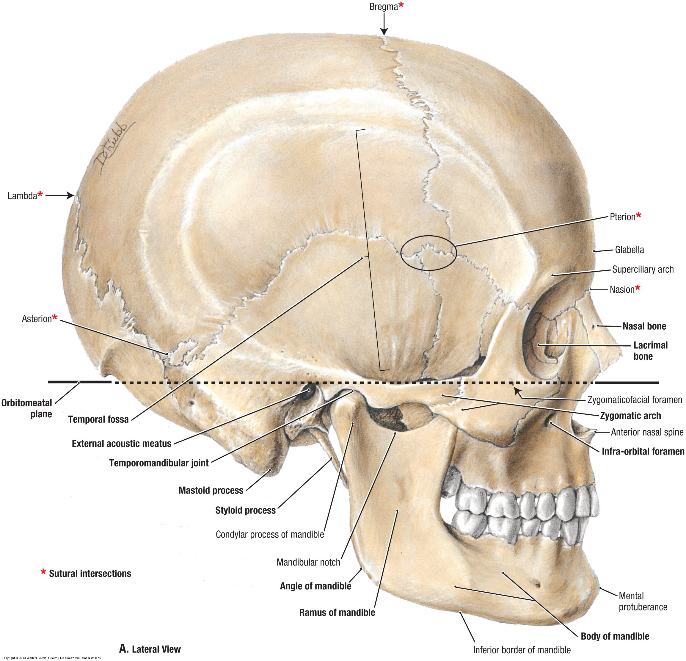
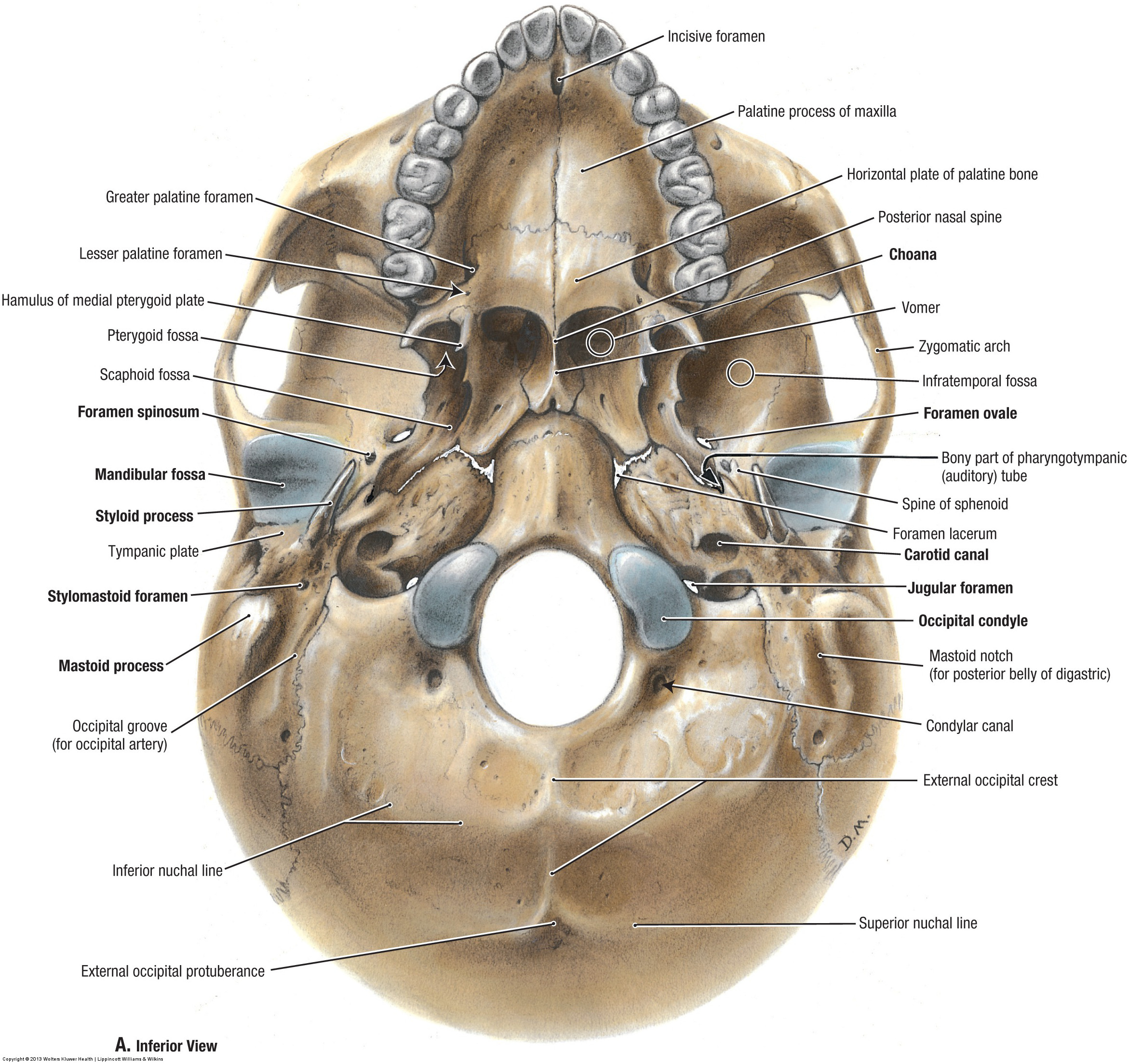
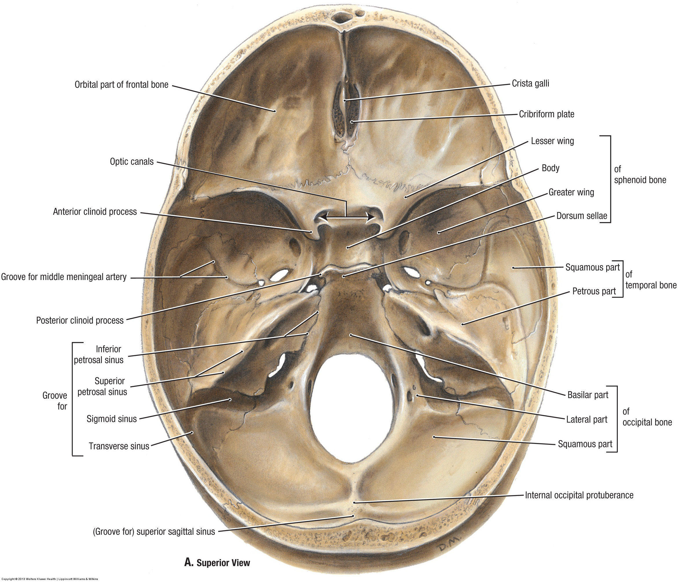
Grant’s Atlas of Anatomy, 14th ed. © 2013 Wolters Kluwer Health | Lippincott Williams & Wilkins
This next set of figures comes courtesy of OpenStax, Anatomy & Physiology. Here the bones are coloured to help you differentiate between them. Click each image to enlarge.
“Download for free at http://cnx.org/contents/14fb4ad7-39a1-4eee-ab6e-3ef2482e3e22@8.24.”
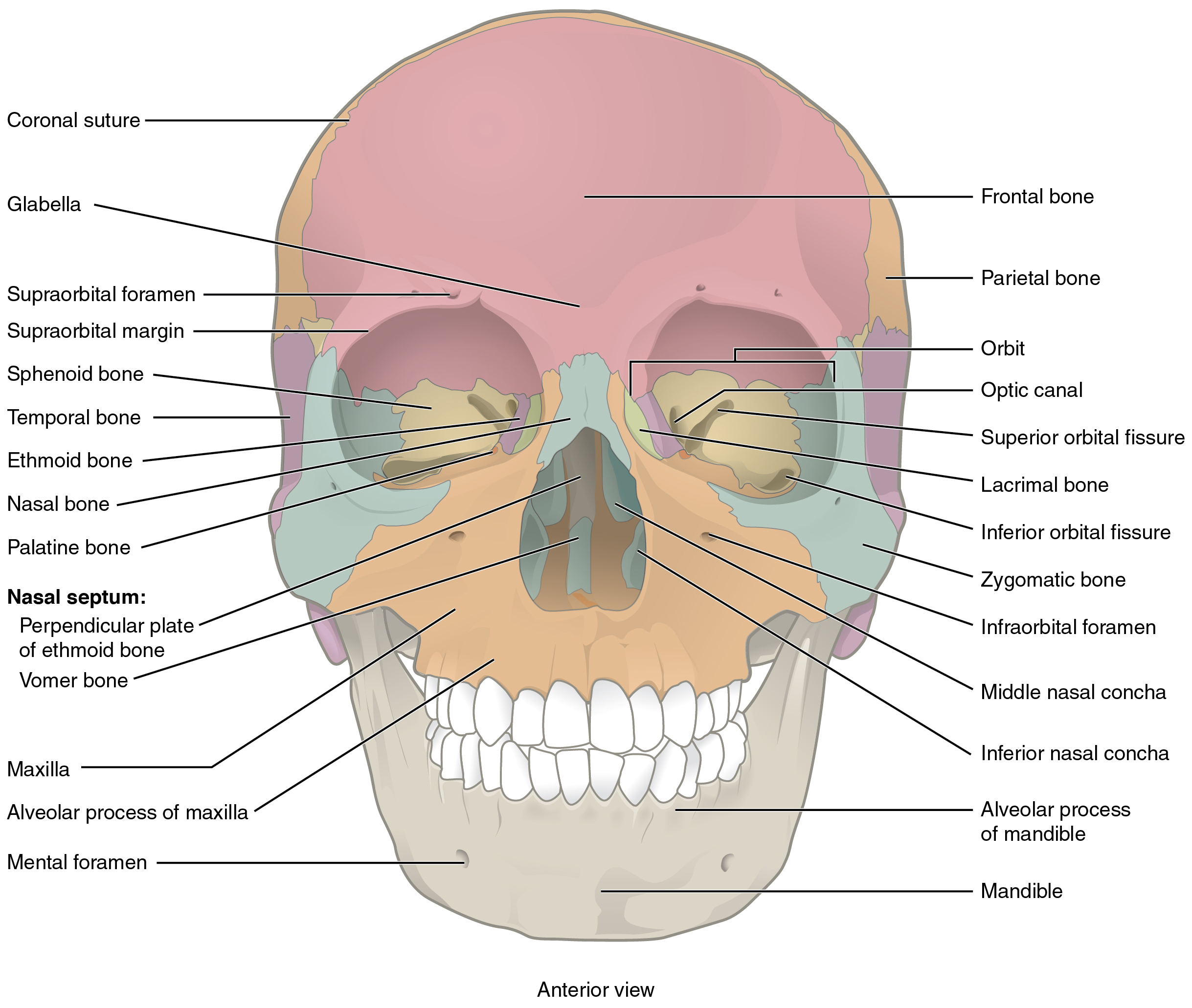
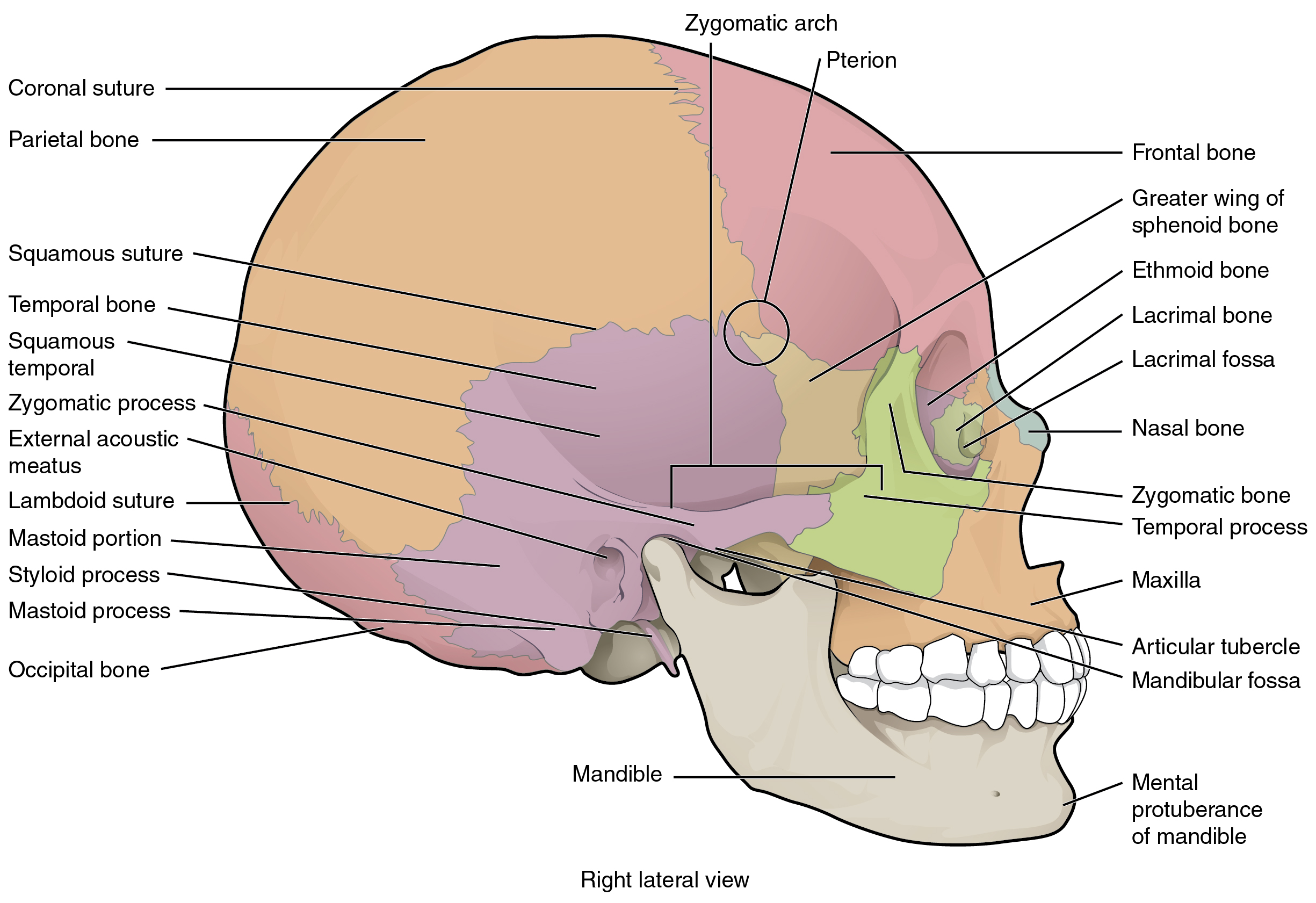
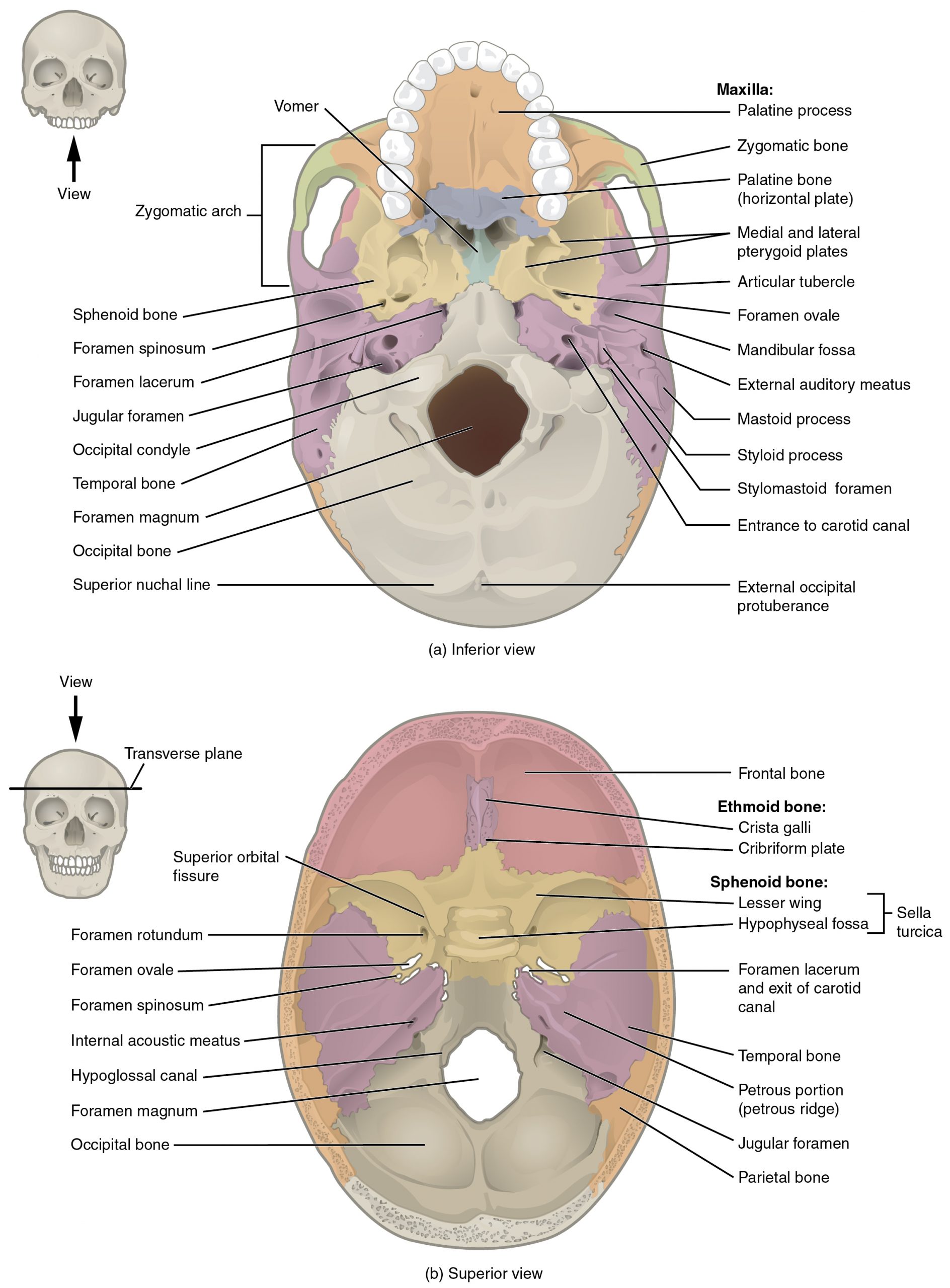
Copyright © 1999-2016 Rice University, CC BY 4.0
“But none of these figures look like the real thing,” you say??
If you don’t own your own copy of Rohen’s Color Atlas of Anatomy (you should!!), here’s a cheeky link to a PDF copy: [click here for beautiful, labelled photos of human anatomy!]
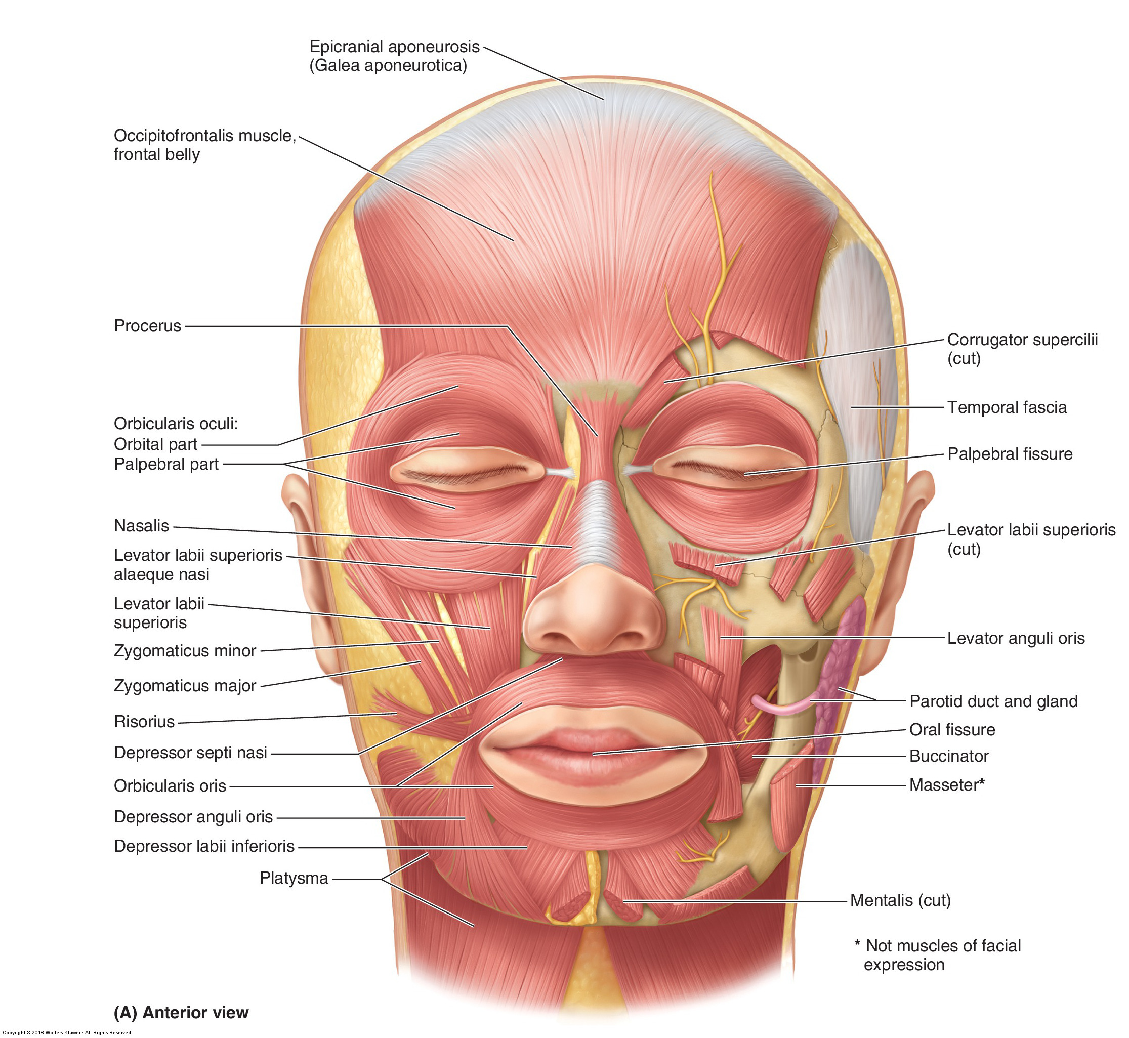
2. Muscles of Facial Expression & Muscles of Mastication
Next, let’s consider the skeletal muscles in this region. Facial muscles can be broadly categorized into 2 groups according to their function:
(a) facial expression or mimetic muscles [Greek: mimētikos = “imitative”]
(b) muscles of mastication [Latin: masticationem = “to chew”]
You will not be expected to know every facial muscle for this course (those included in your lab manual are marked with a black star (★), but here is a complete list for interest’s sake
Facial Expression
★ Occipitofrontalis
★ Orbicularis oculi
☆ Corrugator supercilii
☆ Depressor supercilii
☆ Procerus
☆ Depressor anguli oris
☆ Mentalis
☆ Zygomaticus major
☆ Levator labii superioris
☆ Levator labii superioris alaeque nasi
★ Orbicularis oris
★ Buccinator
☆ Nasalis
☆ Depressor septi nasi
☆ Dilator naris anterior
☆ Depressor labii inferioris
☆ Risorius
☆ Zygomaticus minor
☆ Levator anguli oris
☆ Compressor narium minor
Mastication
★ Masseter
★ Temporalis
★ Medial pterygoid
★ Lateral pterygoid

✓ Facial Expression = innervated by the FACIAL NERVE (CN VII)
✓ Mastication = innervated by the MANDIBULAR branch of the TRIGEMINAL NERVE (CN V3)

Clinically Oriented Anatomy, 8th ed. © Wolters Kluwer
For the muscles of mastication, here are some classic images from the 20th edition of Anatomy of the Human Body by Henry Gray (now Gray’s Anatomy, currently in its 41st edition & edited by Dr. Susan Standring).
*Note that the zygomatic arch, and therefore the masseter, has been removed in both images. The masseter attaches to the lateral surface & ramus of the mandible, extending superiorly and anteriorly to the zygomatic arch. See if you can locate it on the lateral view above (hint: it’s in grey since it’s not a muscle of facial expression)*


These images are in the Public Domain
Interesting fact:
These illustrations were actually by Henry Vandyke Carter, who rarely receives due recognition for his contributions. Henry Gray in fact demanded that the font size be reduced for the illustrator’s name on the title page of Gray’s Anatomy, and Henry Vandyke Carter’s name was removed entirely by the 17th edition in 1909.
3. Meninges of the Brain & Spinal Cord
The brain and spinal cord are encased in 3 protective layers collectively called meninges. If this term sounds familiar you may be thinking of meningitis, which translates to “inflammation (Latin: -itis) of the meninges”]. The 3 layers in question are as follows:
◆ The dura mater [literally translated from Latin = “hard mother”] is the outermost layer. It is a membrane of dense, collagen-rich connective tissue. Around the brain there are 2 layers of dura mater—periosteal (superficial) and meningeal (deep)—between which the dural venous sinuses are located (see Section 4)
◆ The arachnoid mater [Greek: arakhnoeidēs = “cobweb-like”) is the second meningeal layer, made of loose connective tissue. The arachnoid mater has a membranous top layer under which sit irregular trabeculae (columns of loosely-arranged connective tissue). The space under the top membranous layer is known the subarachnoid space and is filled with cerebrospinal fluid (see Section 6). This translucent layer spans across the lumps & bumps (gyri & sulci) of the brain with blood vessels travelling beneath
◆ The pia mater [literally translated from Latin = “tender mother”] is the final layer of meninges. The pia mater closely adheres to the surface of the brain & spinal cord, following all of the irregularities, projections, and grooves. This layer cannot be removed without damaging the underlying brain tissue.

Here’s another artist’s rendition of the meningeal layers, including some additional details about extracranial layers & types of cranial hemorrhage (from Grant’s Atlas of Anatomy, 14th edition).
Notice the arachnoid granulations, projecting from the arachnoid mater into the superior sagittal sinus (see Section 4).
*NOTE: You will not be tested on any topics that are not covered in the lab manual; however some of this ‘extracurricular’ information may be of interest for future clinical practice*

The meningeal layers around the brain continue down the central canal of the spinal column to protect the spinal cord. Below is a rendering from the Brain-inter-atlas—an open-access neuroanatomy atlas developed by teachers at UCLouvain.

How do the meninges differ between the brain and the spinal cord? I’m glad you asked! There are a few clear distinctions of which you should be aware.
◆ In the spinal cord, the arachnoid mater closely lines the internal aspect of the dura mater that forms the dural sac. There is also a distinct space between the dura mater and the vertebra known as the epidural space (in the brain, the periosteal layer of dura mater sits flush with the cranium). The dural sac continues down the spinal canal to approximately the S2 level
◆ In addition to surrounding the spinal cord itself, the pia mater in this region includes lateral extensions called denticulate ligaments [Latin: denticulatus = “small tooth-like”]. There is also a vertical elongation of the pia mater from the base of the conus medullaris (termination of the spinal cord @ ~L1/L2) to the coccyx called the filum terminale [Latin: filum = “thread” + terminalis = “final/terminal”]
Here are a couple of beautiful drawings by Robert Heinrich Johannes Sobotta, a German anatomist best known for his anatomy textbook Atlas der deskriptiven Anatomie des Menschen (now the Sobotta Atlas of Human Anatomy). Click each image to enlarge.
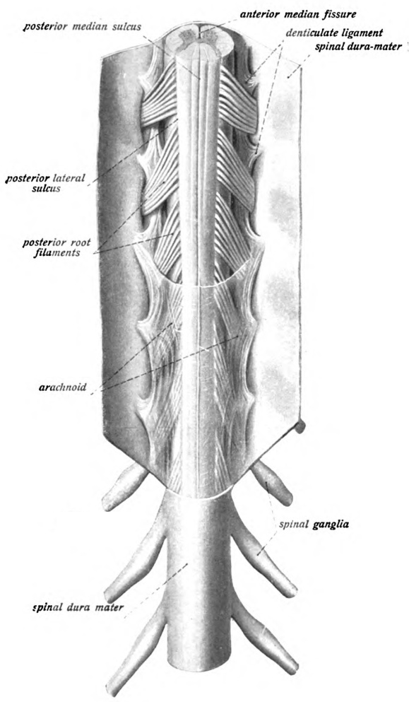
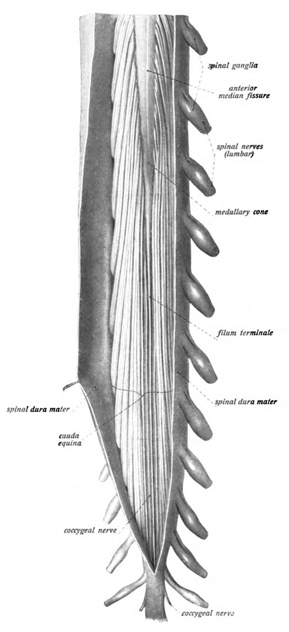
These images are in the Public Domain
I’ve included a few more heavily annotated colour figures from Grant’s Atlas of Anatomy, 14th ed. for your reference. Click each image to enlarge.
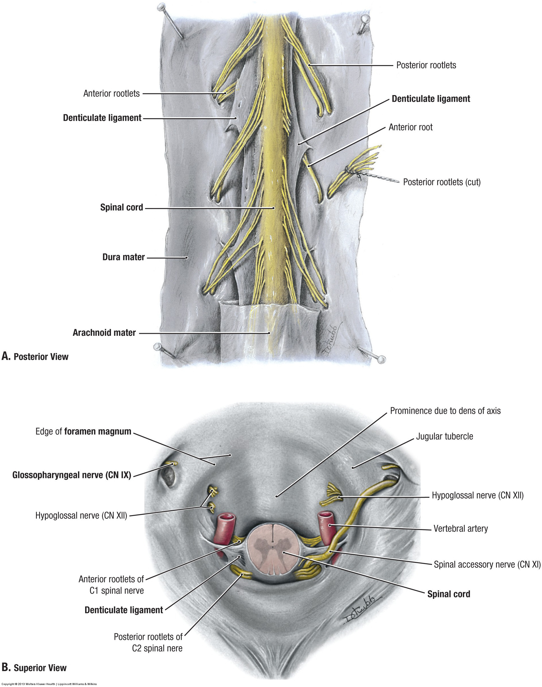
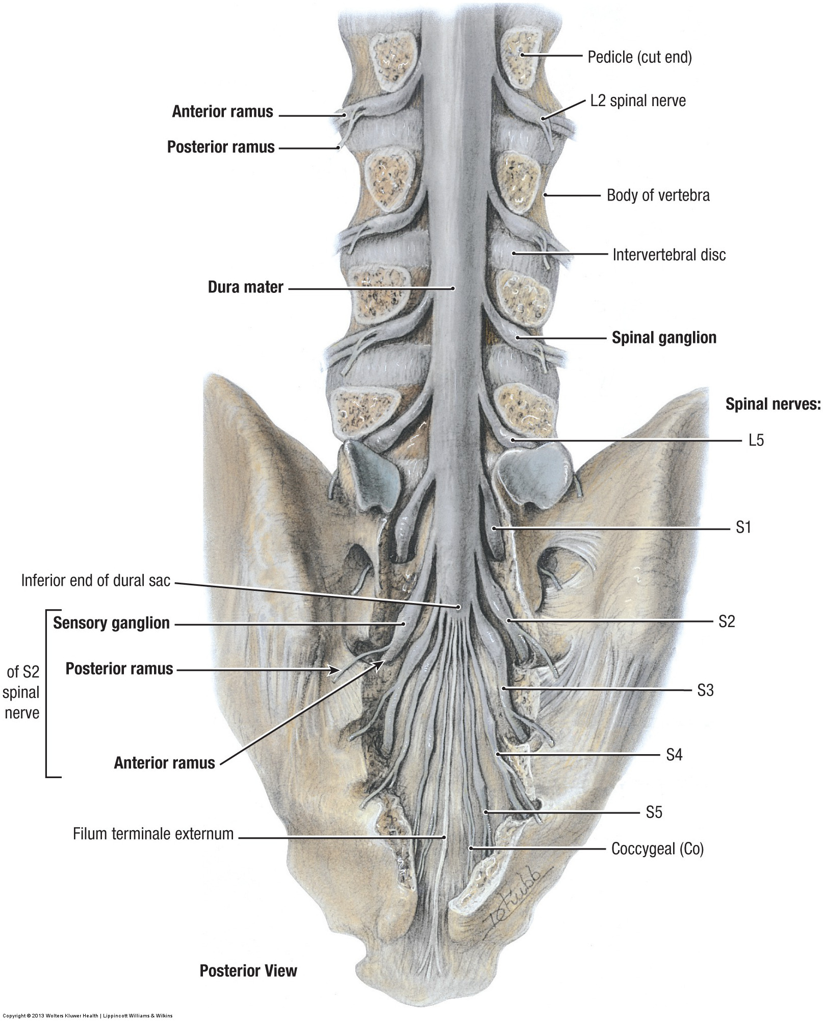
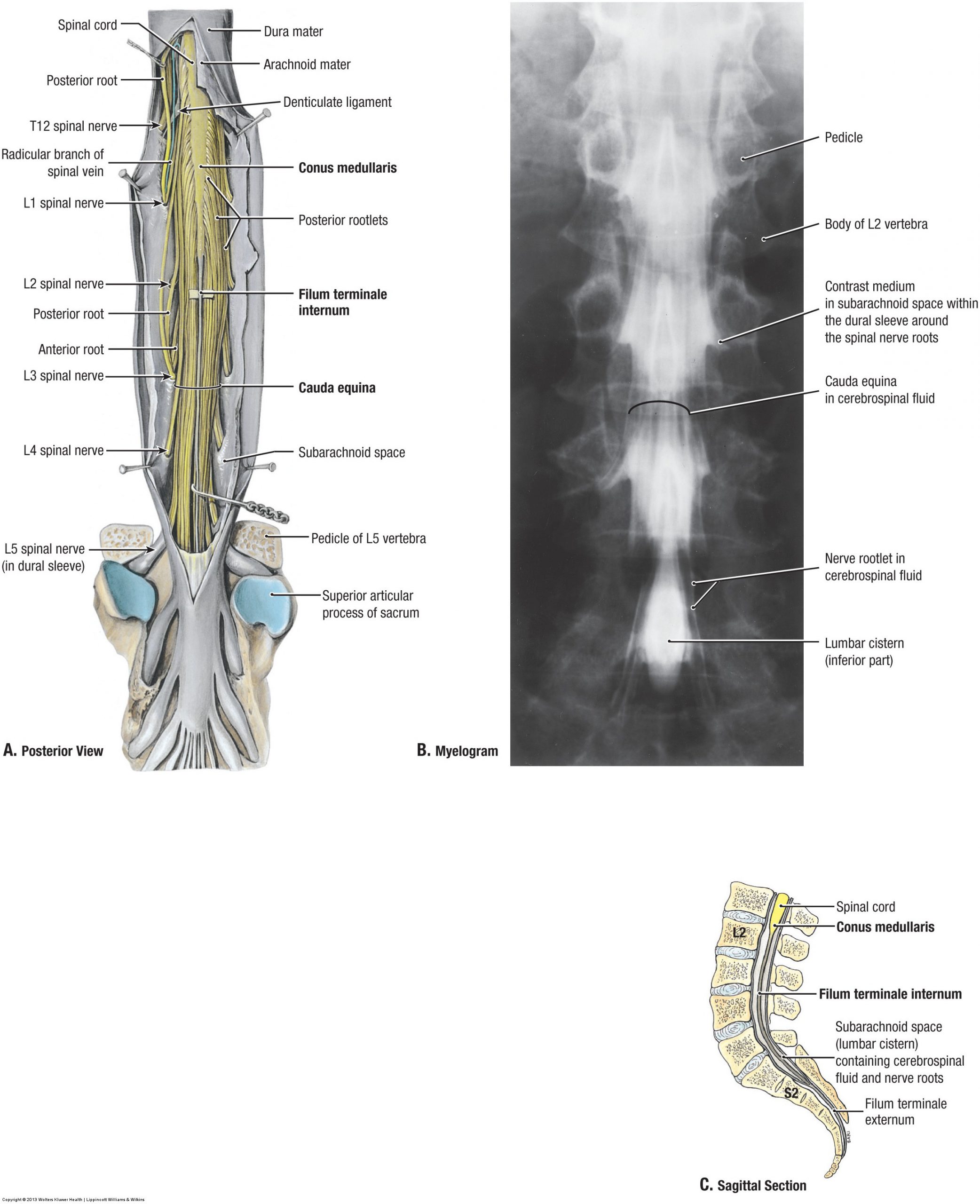
Grant’s Atlas of Anatomy, 14th ed. © 2013 Wolters Kluwer Health | Lippincott Williams & Wilkins
4. Venous Sinuses
Recall that there are two layers of dura mater surrounding the brain: a periosteal layer (on the internal aspect of the skull) and a meningeal layer (superficial to the arachnoid & pia mater). It is between these layers that the dural venous sinuses are situated.
Before we get into the venous sinuses, there are two other structures of which you will need to be aware: the falx cerebri & the tentorium cerebelli. These are known as dural reflections or dural folds because they are formed by the inner meningeal layer of dura mater protruding into the cranial cavity as sheet-like projections. Essentially this results in a double layer of meningeal dura mater, tightly fused together except for where venous sinuses pass through.
◆ The falx cerebri [Latin: falx = “sickle” + cerebrum = “brain”] is a curved (sickle-shaped) dural reflection that separates the left & right hemisphere, protruding into the median longitudinal fissure
◆ The tentorium cerebelli [think of it as a tent for the cerebellum] is a dural reflection that separates the posterior part of the cerebrum from the cerebellum
Here’s a T1-weighted coronal MRI highlighting the outline of the dura mater (including the dural reflections) & a couple of the venous sinuses we are about to discuss. Full open-access article by Iacono et al. (2015) available online here: https://doi.org/10.1371/journal.pone.0124126.

Now that we’ve established where the venous sinuses are located with respect to the cranial meninges it’s time to take a closer look. This unique network of veins is responsible for drainage of blood from the cranial cavity, primarily into the internal jugular veins, bilaterally. These veins (esp. the superior sagittal sinus) also receive cerebrospinal fluid (CSF) from the subarachnoid space, facilitating the rapid systemic turnover of CSF (~3-4 times/day).
There are a couple of interesting distinctions between the venous sinuses and most other veins in the body:
✓ They are *not* accompanied by a set of arteries with similar names & parallel trajectories
✓ They are valveless, allowing for bidirectional flow of blood
The cranial cavity is a complex 3D space. It is perhaps best to appreciate the venous sinuses in a (mostly) empty skull before considering its relationships with all of the other tissues! Take a look at the illustration below (artist: unknown) to see the dural venous sinuses in relation to the dural reflections discussed earlier.

Check out the above illustration (artist unknown) and accompanying flowchart (made by yours truly!)
Still struggling to picture it in 3D? Watch this brilliant little animation by Anatomage illustrating the relationship relationships between the cranial veins and the brain.
"Veins of the Brain" created by Anatomage Table (Direct link: click here)
5. General View of the Brain
This week, we will start by defining and identifying a handful of subdivisions and major features within the brain. To thoroughly appreciate the subdivisions of the brain, one must consider the embryological development of the brain.
The neural tube (embryonic precursor to the central nervous system) differentiates into 3 primary divisions: prosencephalon (forebrain), mesencephalon (midbrain), & rhombencephalon (hindbrain).
◆ The forebrain later differentiates into the telencephalon (cerebrum) & the diencephalon (thalamus, hypothalamus, epithalamus, & subthalamus)
◆ The midbrain does not further subdivide. It ultimately develops into a collection of structures surrounding the cerebral aqueduct (see Section 6), including essential motor nuclei & sensory relay centres. We will explore this area of the brain in cross-section later in the course!
◆ The hindbrain differentiates into the metencephalon (pons), the myelencephalon (medulla oblongata), & the cerebellum (Latin: diminutive of cerebrum = “little brain”).

Now let’s translate this to the adult brain. Here’s what these subdivisions look like once they are fully developed:


Focus on E. and F. in the figure from Grant’s Atlas of Anatomy above.
*Lab 2 will cover detailed structures and landmarks associated with the brain.*
6. Ventricles & Cerebrospinal Fluid (CSF)
The final topic for Lab #1 is the ventricles of the brain. This network of cavities contains cerebrospinal fluid (CSF): a transparent, colourless fluid that plays an essential role in protecting the brain (think of it like a fluid-filled cushion), waste removal, and more. CSF can be found around the brain and spinal cord in the subarachnoid space (review Section 3).
CSF is produced by the choroid plexus, found primarily in the lateral ventricles & fourth ventricle). Enough CSF is produced each day to replace the entire systemic volume ~3-4 times per day, necessitating an efficient system for resorption. Recall the arachnoid granulations (Section 3)—these outpouchings of the arachnoid mater serve to absorb CSF and return it to the blood stream. An imbalance between production and resorption resulting in too much CSF will lead to hydrocephalus [Greek: hudro- + kephalē = ” water head”]. This may be due to (a) overproduction, (b) reduced absorption, (c) obstruction of flow, or a combination of factors.
Below you will find a picture of choroid plexus in the lateral ventricle (coronal section), as well as an illustration of the ventricles and a handy flowchart!


Need a quick review?
Check out this 2-minute recap by Dr. Marc Dingman (creator of Neuroscientifically Challenged) to review the circulation of cerebrospinal fluid in the ventricles of the brain!
"The Ventricles" created by Neuroscientifically Challenged (Direct link: click here)
Miscellaneous Study Materials 🧠
Sporcle Quiz: Fun with Foramina—Who Goes There Edition
Made by Mikaela Stiver (yours truly!)
Adventures in Neuropathology: Twitter Post & Video
How many structures & functions can you name?
This post is from the Adventures in Neuropathology Twitter page (@AdvNeuroPath). Answers are available in the following video by Dr. Andrea Gilbert: Click here!
Beautiful example of normal neuroanatomy. Can you identify all of the normal structures? Annotated photo will be posted tomorrow pic.twitter.com/ZiIbE6Xaz1
— Adventures in Neuropathology (@AdvNeuroPath) April 4, 2020
UBC: 3D Model of the Brainstem + Cerebral Arterial Circle
Here’s a fantastic annotated model of the brainstem + cerebral arterial circle (eponym: circle of Willis) by Ishan Dixit, Connor Dunne, and Curtis J. Logan for UBC Medicine – Educational Media. Turn off the annotations & test yourself!
Neuropathology Illustrated Quizzes: Drs. Townsend & Klatt
All content is subject to Copyright © 1999,2001 by Jeannette J. Townsend MD, Department of Pathology, University of Utah, Salt Lake City, Utah, USA, and by designated contributors. All rights reserved worldwide.
Soton Brain Hub: General Neuroanatomy Quizzes
Soton Brain Hub was developed by students, staff, & alumni of the University of Southampton. It is continually updated, including a YouTube channel & Instagram account!
These quizzes & others are on the Soton Brain Hub webpage: www.sotonbrainhub.co.uk
Soton Brain Hub "General Quiz 1", available on www.sotonbrainhub.co.uk